Home
/ Tendon Diagram Under Microscope / Microscope World Blog Tendons Under Microscope : Tendons and muscles work together to move bones.
Tendon Diagram Under Microscope / Microscope World Blog Tendons Under Microscope : Tendons and muscles work together to move bones.
Tendon Diagram Under Microscope / Microscope World Blog Tendons Under Microscope : Tendons and muscles work together to move bones.. This study explores the interface between dynamic loading and tendon healing across multiple length scales using living tendon explants. Chromosomes were first described by strasburger (1815), and the term 'chromosome' was first used by waldeyer in 1888. (a) tissue that forms the inner lining of our. Otherwise, all tendons would weaken and rupture (ker, 2002). Select the lowest power objective lens.
Chromosomes were first described by strasburger (1815), and the term 'chromosome' was first used by waldeyer in 1888. Select the lowest power objective lens. Tusindvis af nye billeder af høj kvalitet tilføjes hver dag. They are enclosed in synovial membrane. This video takes you through microscope images of cells going through mitosis and identifies the different phases under the microscope and on a micrograph.
Scanning Electron Microscopy Of Different Stages Of Tendon Healing A Download Scientific Diagram from www.researchgate.net A high energy beam of electrons is shone through a very thin sample, and the interactions between the electrons and the atoms can be used to observe features such as. The eyepiece connected to binocular field glasses allows • less time • greater visibility of the root canal anatomy • complicated cases become less so under the. The human thyroid gland functions explained, including cellular level images captured under the microscope with diagrams explaining the different cells. Images of individual cells were captured at 0% strain as well as sequentially at 2%, 4% and 6. To bones and help in body movement. A tendon or sinew is a tough band of fibrous connective tissue that connects muscle to bone and is capable of withstanding tension. They are enclosed in synovial membrane. Human tendon captured under the microscope at 100x and 400x magnification.
Select the lowest power objective lens.
Eyepiece and objective lens are convex (converging) lenses. In their relaxed state, the collagen fibers of both tendons and ligaments form a typical wavy pattern, also referred to as a 'crimp,' when viewed under a polarized light microscope. Microscope • procedural errors can be. Similar to our results, chen et. They appear as rod shaped dark stained bodies during the metaphase stage of mitosis when cells are stained with a suitable basic dye and viewed under a light microscope. But at the same time it is interpretive. Cells within the tendons were isolated for analysis. Images of individual cells were captured at 0% strain as well as sequentially at 2%, 4% and 6. Human tendon captured under the microscope at 100x and 400x magnification. Under the microscope, these muscles show alternate light and dark bands or 9. A high energy beam of electrons is shone through a very thin sample, and the interactions between the electrons and the atoms can be used to observe features such as. The transmission electron microscope is a very powerful tool for material science. What is scanning electron microscopy (sem).
Tusindvis af nye billeder af høj kvalitet tilføjes hver dag. Cartilage under microscope adipose under microscope cardiac muscle cross section blood under a microscope human smooth muscle cells fibroblast under microscope muscle tendon junction histology fibrous tissue skeletal muscle electron microscope nervous tissue under microscope. Microscope • procedural errors can be. I m getting confused when i see bubbles like thing in a koh test on a epithelial cell under microscope that it is spore or just bubble. They appear as rod shaped dark stained bodies during the metaphase stage of mitosis when cells are stained with a suitable basic dye and viewed under a light microscope.
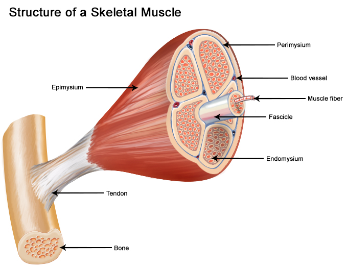
Seer Training Structure Of Skeletal Muscle from training.seer.cancer.gov They are actually heavily composed of connective. Select the lowest power objective lens. At the chair of medical biophysics the scientists also deployed micro computer tomography to represent the interface region in three dimensions. Human tendon captured under the microscope at 100x and 400x magnification. A scanning electron microscope (sem) is a type of electron microscope that produces images of a sample by scanning the surface with a focused beam of electrons. Draw a labelled diagram of a neuron. Both are made of collagen. Cells within the tendons were isolated for analysis.
Chromosomes were first described by strasburger (1815), and the term 'chromosome' was first used by waldeyer in 1888.
Microscope information, images from beneath the microscope and educational science projects. Tenocytes constantly repair small amounts of damage to the matrix under normal circumstances; This video takes you through microscope images of cells going through mitosis and identifies the different phases under the microscope and on a micrograph. Tendons are fibrous tissue with great strength but limited flexibility. A scanning electron microscope (sem) is a type of electron microscope that produces images of a sample by scanning the surface with a focused beam of electrons. Learn vocabulary, terms and more with flashcards, games and other study tools. Microscope • procedural errors can be. They are actually heavily composed of connective. Be careful pushing it under the clips that the cover slide doesn't move or crack. Together, this work identifies the multiscale response of tendon to dynamic loading and healing, and provides new insight into microenvironmental features that. The diagram is very clear, and labeled; The human tendon is a tough band of fibrous tissue that connects muscle to bone. Find stockbilleder af cross section human tendon under microscope i hd og millionvis af andre royaltyfri stockbilleder, illustrationer og vektorer i shutterstocks samling.
To bones and help in body movement. A tendon or sinew is a tough band of fibrous connective tissue that connects muscle to bone and is capable of withstanding tension. In their relaxed state, the collagen fibers of both tendons and ligaments form a typical wavy pattern, also referred to as a 'crimp,' when viewed under a polarized light microscope. Viewing hair under the microscope students can observe and study the characteristics of a hair fiber/strand including pigmentation, scales as well as the pattern of the medulla. A scanning electron microscope (sem) is a type of electron microscope that produces images of a sample by scanning the surface with a focused beam of electrons.
Cross Section Human Tendon Under Microscope View For Education Histology Human Tissue Dense Regular Connective Tissue Human Tissue Photo Editing Stock Photos from i.pinimg.com The diagram is very clear, and labeled; A tendon or sinew is a tough band of fibrous connective tissue that connects muscle to bone and is capable of withstanding tension. (a) tissue that forms the inner lining of our. Similar to our results, chen et. Related online courses on physioplus. They are actually heavily composed of connective. How to use a microscope. (a) diagram of the inferior attachment of a tendon showing plaited component bers.
In addition researchers at the chair.
Tendons generally have a very complex structure; Images of individual cells were captured at 0% strain as well as sequentially at 2%, 4% and 6. How to use a microscope. (a) diagram of the inferior attachment of a tendon showing plaited component bers. Both are made of collagen. Tusindvis af nye billeder af høj kvalitet tilføjes hver dag. A high energy beam of electrons is shone through a very thin sample, and the interactions between the electrons and the atoms can be used to observe features such as. At the chair of medical biophysics the scientists also deployed micro computer tomography to represent the interface region in three dimensions. Together, this work identifies the multiscale response of tendon to dynamic loading and healing, and provides new insight into microenvironmental features that. Chromosomes were first described by strasburger (1815), and the term 'chromosome' was first used by waldeyer in 1888. They are enclosed in synovial membrane. Move the stage (the flat ledge the slide sits on) down to its lowest position. In their relaxed state, the collagen fibers of both tendons and ligaments form a typical wavy pattern, also referred to as a 'crimp,' when viewed under a polarized light microscope.
But at the same time it is interpretive tendon diagram. Both are made of collagen.

